DNA Amplification
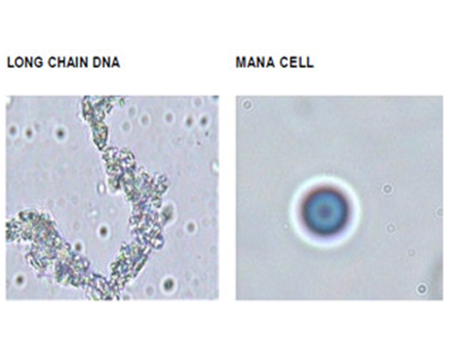
When patients plasma containing WBCs is treated with Manaitleadsto DNA genesis and Manacell formation,simultaneously within seconds, which can be seen using inverted microscope under white light as scattered particles of irregular shape and Manacells which are regularly round uniform with colors of the vibgyor the nucleus being red and enlarged, with the surface markers not seen due to a thickened cell wall in color blue.
On addition of Mana to patients’ plasma the huge chains of DNA are formed with varying sizes which is over 20 to 50 microns in size as compared to the Manacell which is close to 10 microns. And these chains of DNA and Manacells when viewed at higher magnification under white light gives an appearance of long chains of DNA and smooth round polychromatic Manacells. The pleats of the naked DNA chains with no cell wall or nuclear membrane can be seen and this is confirmed using the DNA stains and sequencing done at later points in this study.
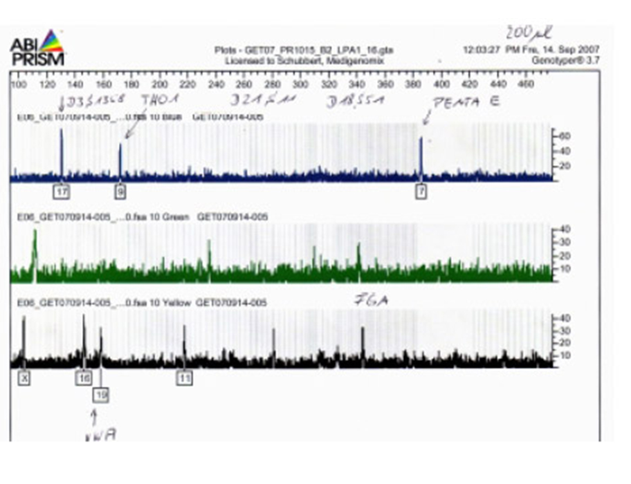
DNA ALLELE MARKERS SHOWING POSITIVE FOR THE DNA GENERATED
The DNA thus generated was sent to an independent laboratory in Europe where allele markers were used to identify it as being an uncontaminated human DNA from the plasma of a female patient, since the allele markers were positive for both big and small chain DNAs there was no denaturing of the protein.
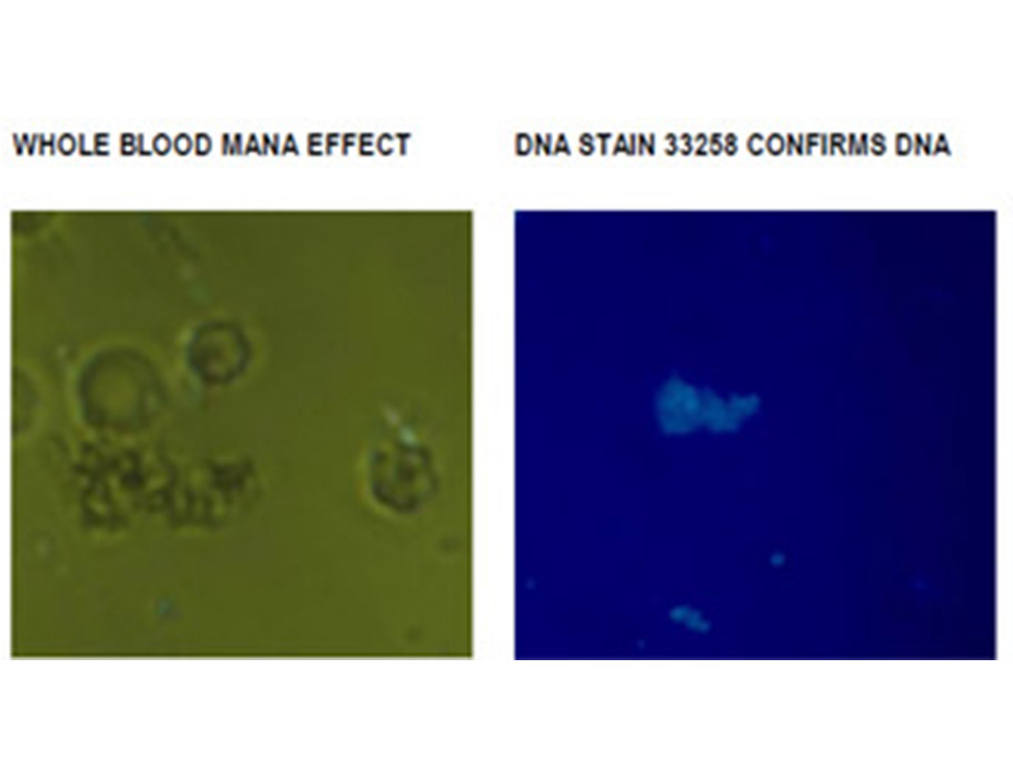
On addition of Mana to whole blood the generation of Manacells and DNA takes place. Which is confirmed with DNA staining dye HOECHST 33258 that gives blue fluorescence when excited under ultraviolet light. Under normal green light and high magnification once again the contour of the Manacells can be seen to be uniformly round, since green light was used here the vibgyor colors are not noticed. There are more than one DNA particles in every field of the specimen under study.